Book a Service
1800 120 51600
Tollfree Helpline

What is Fetal Medicine?

Fetal medicine, also known as maternal-fetal medicine (MFM) is a subspecialty of obstetrics that focuses on the health of the mother and fetus before, during, and after pregnancy. Fetal medicine has advanced in many ways to monitor and manage maternal and fetal health, including through technological innovations and medical breakthroughs.

Who are Fetal Medicine Specialists

Fetal Medicine Specialists are obstetricians and gynaecologists with additional training in managing high-risk pregnancies. They work with pregnant women and their primary obstetricians to address complications and optimize the health of both the mother and the fetus. Petals Imaging offers one of the best Fetal medicine Services in Kolkata. Learn why?

Role of Fetal Medicine in Obstetrics

  • Advanced Ultrasound diagnosis: Fetal ultrasound has revolutionized prenatal care by providing detailed information about fetal growth, anatomy, and well-being throughout the pregnancy. This helps in the  early detection of abnormalities, and advanced technologies like 3D/4D ultrasounds can provide even more detailed information.
  • Diagnostic tests: Maternal-fetal medicine specialists use various diagnostic tests and monitoring procedures to assess fetal health, including fetal echocardiography, genetic screening tests, and comprehensive maternal health assessments.
  • Monitoring equipment: Equipment used to monitor fetal cardiac activity, maternal contractions, amniotic fluid volume, and vascular resistance includes external fetal monitors, intravenous oxytocin, vibroacoustic stimulators, and ultrasound with Doppler velocimetry.
 

List of Scans During Pregnancy:

Here’s a list of typical scans performed during pregnancy, arranged chronologically by weeks:

First Trimester (Weeks 1–12):

  1. Early Pregnancy Scan (4–6 weeks):
    • Identifying Gestational Sac and location of the pregnancy. Sometimes it may be difficult to determine if the pregnancy is viable at this stage.
    • Addresses concerns like vaginal bleeding, abdominal pain, or suspected miscarriage.
  2. Dating and Viability Scan (6–8 weeks):
    • Confirms pregnancy.
    • Checks for fetal heartbeat, number of fetuses, and estimated gestational age.
  3. Nuchal Translucency (NT) Scan (11–14 weeks):
    • Part of the first-trimester screening for chromosomal abnormalities like Down syndrome.
    • Measures the fluid at the back of the baby’s neck.

Second Trimester (Weeks 13–28):

  1. Anatomy (Anomaly) Scan (18–22 weeks):
    • Detailed examination of the baby’s organs, limbs, and spine.
    • Assesses placenta position, amniotic fluid levels, and fetal growth.
  2. Cervical Length Scan (16–24 weeks, if needed):
    • Checks for cervical insufficiency in high-risk pregnancies.

Third Trimester (Weeks 29–40):

  1. Growth Scan (28–32 weeks):
    • Monitors baby’s growth, position, and amniotic fluid levels.
    • Checks for complications like restricted growth.
  2. Doppler Ultrasound (32–36 weeks, if needed):
    • Evaluates blood flow in the umbilical cord and fetal vessels to ensure proper oxygen and nutrient delivery.
  3. Position/Wellbeing Scan (36–40 weeks):
    • Confirms the baby’s position (head-down, breech, etc.).
    • Assesses placenta, fluid levels, and overall health.

Additional/Optional Scans:

Important screenings done in conjunction with maternal-fetal medicine include:

  • Alpha-fetoprotein testing
  • Amniocentesis
  • Pre-eclampsia Screening
  • Chorionic villus sampling (CVS)
  • Down syndrome screening
  • Multiple marker screening (MMS)
  • Neural tube defect screening
  • Fetal cardiac anomaly screening
  • Non-stress/contraction stress tests (fetal heart rate testing)

Understanding the importance and Purpose of Fetal Medicine in ensuring a Safe Pregnancy

  • Assessing structural abnormalities
  • Evaluating Fetal health
  • Assessing Maternal health
  • Prenatal Screening for Genetic abnormalities
  • Fetal Diagnosis

Ultrasound scan to assess structural abnormalities

  • Early anomaly scan (12-16 weeks): A detailed ultrasound is performed between 12-16 weeks of pregnancy to identify major fetal abnormalities and assess overall fetal development.
  • Second-trimester anomaly scan or Level-II scan: A comprehensive ultrasound around 18-20 weeks to evaluate the baby’s anatomy and screen for structural or genetic abnormalities.
  • Multiple Pregnancy Scan: This scan monitors the development and health of each fetus during multiple pregnancies, assessing growth and identifying potential complications.
Second trimester scans

Ultrasound scan to Evaluate Fetal health

Used to assess the overall health and development of the fetus, including growth, position, and amniotic fluid levels.
  • Fetal viability scan: Performed in early pregnancy to confirm fetal heartbeat and ensure the pregnancy is progressing normally.
  • Fetal well-being scan: A detailed scan that assesses fetal growth, position, movements, and amniotic fluid to ensure the baby is healthy.
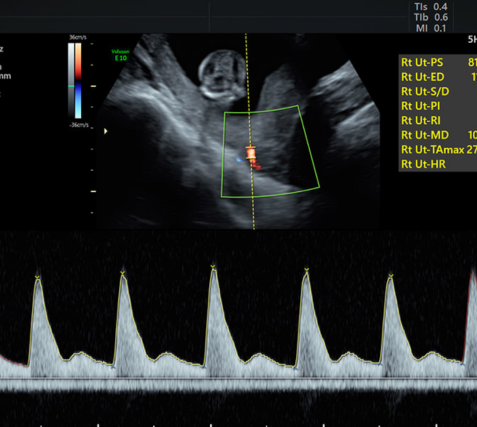
  • Doppler scan: An ultrasound that measures blood flow in the fetus’s vessels, helping assess oxygen and nutrient supply.
  • Fetal echocardiography: Specialized ultrasound to evaluate the fetal heart’s structure, function, and rhythm, helping detect heart defects.
  • Fetal Neurosonogram: A detailed scan focusing on the fetal brain and spinal cord, detecting abnormalities in the nervous system.
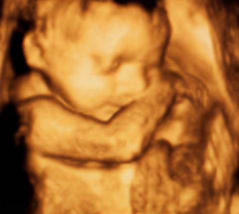
  • 3-D and 4-D Ultrasound: Advanced imaging that provides three-dimensional and real-time moving images of the fetus, enhancing visualization of features.
3D/3D doppler ultasound of fetus. Fetus seen in 3D-Fetal medicine at Petals health

Ultrasound scan to assess Maternal health

  • Pre-eclampsia screening: Screening that uses ultrasound and blood tests to assess the risk of pre-eclampsia, a condition characterized by high blood pressure during pregnancy.
Pre-Eclampsia Screening via ultrasound - checking the blood flow to the uterus.

Non-Invasive Screening Tests:

Tests that evaluate the risk of genetic disorders in the fetus without needing invasive procedures via blood tests and an ultrasound scan at specified weeks of Gestation.
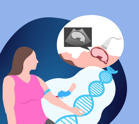
  • First Trimester Screening: A combination of ultrasound and blood tests performed between 11-14 weeks to assess the risk of chromosomal abnormalities like Down syndrome.
  • Second Trimester Screening: A follow-up screening using blood tests and ultrasound to detect genetic abnormalities and evaluate fetal growth.

Invasive Prenatal Diagnosis Procedures:

Fetal Diagnostic procedures, including Chorionic Villi Sampling, Amniocentesis and Fetal Intervetions like Fetal reduction. Only Certified Genetic Clinics – Ultrasound Clinics with Genetic Diagnosis licences from the government are allowed to do these procedures. These procedures are done by skilled and experienced Fetal Medicine specialists only, as these procedures are slightly invasive and have a slight risk. They are only done as a confirmatory test when primary screening tests show a strong indication of risks to the fetus and pregnancy outcome. Sometimes, Fetal Intervention can be carried out to correct the risks.
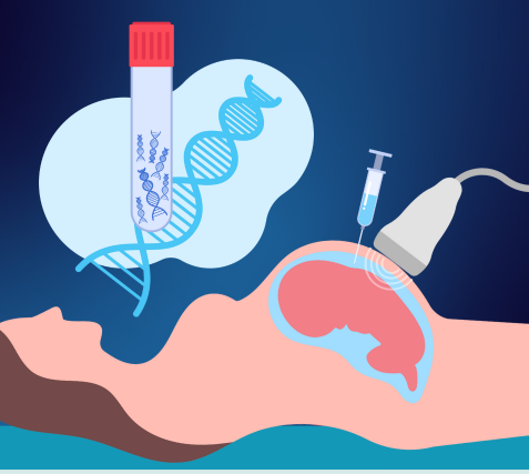
  • Chorionic Villus Sampling: A diagnostic test where a small sample of placental tissue is taken to test for genetic conditions.
  • Fetal Blood Sampling: A procedure to collect a sample of the fetus’s blood to test for infections or genetic abnormalities.
  • Amniocentesis: A procedure where amniotic fluid is sampled to check for chromosomal abnormalities or genetic disorders.
  • Fetal Reduction: A procedure used to reduce the number of fetuses in multiple pregnancies to lower health risks for the mother and remaining fetuses.

Watch Our Videos

Aesthetic procedures aim to enhance one’s physical appearance, boost self-confidence, and promote a sense of well-being.

  • English
  • Bengali
  • Hindi

Understanding Maternal Fetal Medicine and Fetal Ultrasound

Understanding Maternal Fetal Medicine and Fetal Ultrasound

Understanding Maternal Fetal Medicine and Fetal Ultrasound

Understanding

Conclusion

Maternal Fetal Medicine (MFM) is a specialized branch of obstetrics focused on managing complex pregnancies and ensuring the health of both the mother and fetus. This field uses a range of advanced diagnostic tools and scans to monitor fetal development, detect abnormalities, and assess maternal well-being throughout pregnancy. Key scans in MFM include NT scans, second-trimester anomaly scans, fetal viability and wellbeing scans, Doppler studies, and specialized imaging like fetal echocardiography and neurosonograms. Together, these scans provide a comprehensive understanding of fetal health, enabling early detection and management of potential complications for better pregnancy outcomes.


  • Timely completion of these maternal-fetal scans is crucial for the health of both the mother and fetus, as well as for ensuring a positive pregnancy outcome.
  • Early and regular scans allow prompt detection of structural abnormalities, genetic conditions, or complications that may otherwise go unnoticed.
  • By identifying these issues early, healthcare providers can intervene and manage potential risks, reducing the chances of severe complications later in pregnancy or at birth.
  • For the mother, timely scans can help detect conditions like pre-eclampsia, gestational diabetes, or cervical insufficiency, which may pose significant health risks if left untreated.
  • Early detection allows for better management and monitoring, reducing the likelihood of emergency interventions and improving maternal health.
  • For the fetus, scans such as anomaly screenings and Doppler studies can reveal problems with growth, blood flow, or organ development, allowing for targeted treatments or preparations for any necessary interventions post-birth.
  • Monitoring fetal health can also guide decisions regarding delivery timing and method, helping to prevent complications during labour and delivery.

Ultimately, regular and timely scans lead to improved pregnancy outcomes by allowing for early diagnosis, timely treatment, and well-planned care, ensuring the best possible health for both mother and baby.


Explore Other Scans During Pregnancy

Petals Imaging Highlights:

If you are pregnant and looking for a reliable and trustworthy ultrasound centre, book your pregnancy scans at Petals Imaging. We are operational at three locations across Kolkata. Here’s why your scan experience at Petals Imaging will be the best in Kolkata:

  • We have the best radiologist and fetal medicine experts in Kolkata as well as
  • We use the best Women’s and Obstetric Ultrasound Machines – Ge Voluson S6 in all our clinics.
  • Our experts TAKE TIME IN DOING YOUR SCANS  making sure all the fetal parameters are assessed in detail and accurately
  • With their years of experience in performing complex scans and diagnosing difficult conditions, combined with the advanced tools in the ultrasound machine, our Fetal Medicine experts deliver the most reliable Pregnancy Ultrasound reports in Kolkata.
  • Our Clinic staff and Nurses are supportive and take all the possible care to ensure a clean, hygienic and comfortable scan experience.